
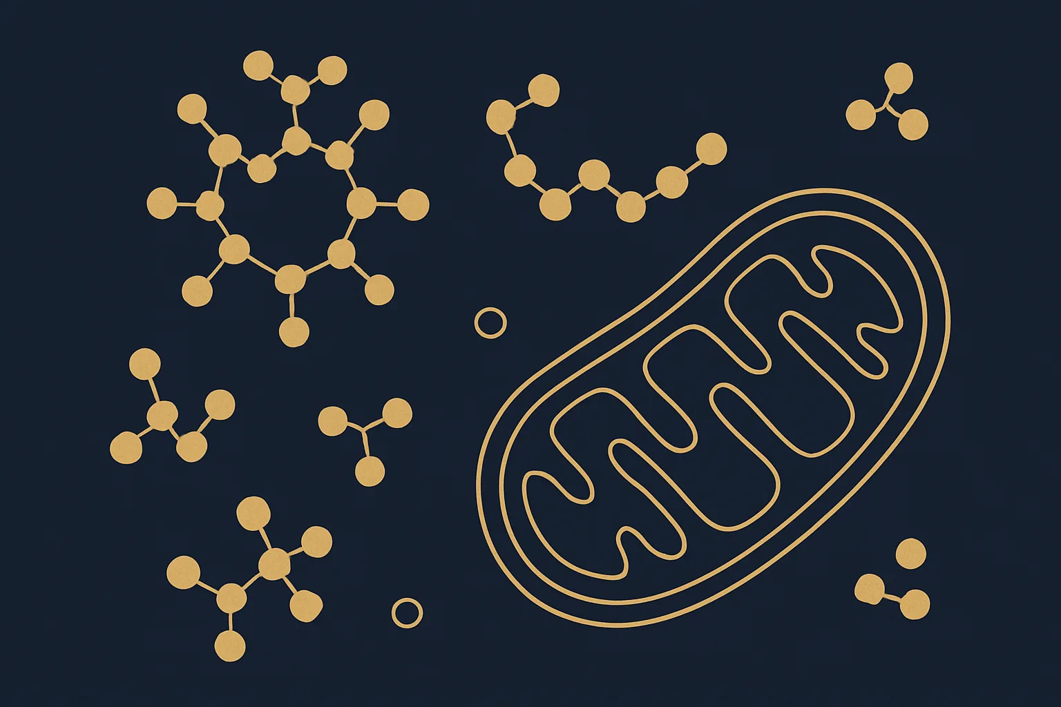
Mitochondrial NAD+ is a functionally distinct pool from cytoplasmic NAD+—not freely exchangeable across the inner mitochondrial membrane (IMM) under basal conditions due to the impermeability of the IMM to charged molecules at physiological pH. Mitochondrial NAD+ is replenished by NMNAT3 (nicotinamide mononucleotide adenylyl transferase 3), the mitochondrial isoform of the NMNAT family, which converts NMN to NAD+ within the mitochondrial matrix using ATP from oxidative phosphorylation. This localized synthesis distinguishes mitochondrial NAD+ homeostasis from cytoplasmic pools (maintained by NMNAT1 in the nucleus and NMNAT2 in the cytoplasm/Golgi). In aged liver mitochondria, NAD+ concentration is reduced approximately 35–45% compared to young animals (isolated mitochondria HPLC), while cytoplasmic NAD+ shows a different decline trajectory—demonstrating the importance of compartment-specific measurement.
Complex I (NADH:Ubiquinone Oxidoreductase) and NAD+ Dependency
Complex I (CI) is the entry point for NADH-derived electrons into the ETC. Its 44-subunit structure includes an NADH-oxidation module that transfers electrons from NADH (generated by TCA cycle dehydrogenases—IDH3, α-KGDH, MDH2, OGDH) to ubiquinone, simultaneously pumping 4H+ per 2e- across the IMM to maintain ΔΨm. CI function requires: (1) available NAD+ as oxidized electron acceptor for matrix dehydrogenases; (2) functional CoQ pool; (3) intact CI structural assembly (14-core subunits + 30 accessory subunits, maintained by assembly factors NDUFAF1-8). Aging disrupts CI through: CI subunit oxidation (particularly NDUFV1 and NDUFB8, susceptible to ROS-mediated carbonylation), CI assembly defects (NDUFAF4 and NDUFAF2 decline in aged muscle), and reduced NADH/NAD+ ratio secondary to NAD+ depletion. Blue native PAGE of aged muscle mitochondria shows approximately 20–30% reduction in CI activity staining versus young controls.
NAMPT Regulation and the Biosynthetic Bottleneck
NAMPT (nicotinamide phosphoribosyltransferase) is the rate-limiting enzyme of the NAD+ salvage pathway and a dual cytoplasmic/secreted protein. Intracellular NAMPT (iNAMPT) generates NMN from NAM + PRPP, while extracellular NAMPT (eNAMPT) has been identified as a signaling cytokine (also called PBEF/visfatin) with NAD+-biosynthetic activity at the cell surface. NAMPT is regulated by: (1) substrate availability (NAM, PRPP); (2) SIRT1-dependent deacetylation that increases NAMPT activity (creating a positive SIRT1-NAMPT feedback loop); (3) NF-κB-mediated transcriptional induction in inflammatory contexts (stress-responsive); and (4) PARP-1/CD38-mediated NAM release, which feeds back to NAMPT substrate but also inhibits NAMPT at high concentrations (product inhibition). In aged animals, the SIRT1-NAMPT positive loop is broken: reduced NAD+ decreases SIRT1 activity, which reduces NAMPT deacetylation, which further decreases NMN production, creating a self-amplifying depletion cycle.
Research Parameters: Measuring Compartment-Specific NAD+ and Targeting the Axis
- Mitochondrial NAD+ measurement: isolate mitochondria by differential centrifugation (600×g then 10,000×g), wash in sucrose buffer, lyse in 1 M HClO₄, neutralize, quantify by HPLC (C18 column, 260 nm UV detection) or enzymatic cycling assay—distinguish from total cell NAD+ which conflates compartments
- Complex I activity: NADH:ubiquinone oxidoreductase spectrophotometric assay (340 nm, rotenone-sensitive fraction); Blue Native PAGE complex-in-gel activity staining
- NAMPT activity: NMN production from NAM + PRPP by HPLC measurement; NAMPT protein by western blot (antibody specificity critical—distinguish from PBEF); eNAMPT in plasma by ELISA
- Purity standard: HPLC >99%, lot-traceable CoA for all NAD+ precursors
All research compounds described are for laboratory and preclinical research use only. They are not approved for therapeutic, diagnostic, or clinical applications in humans.